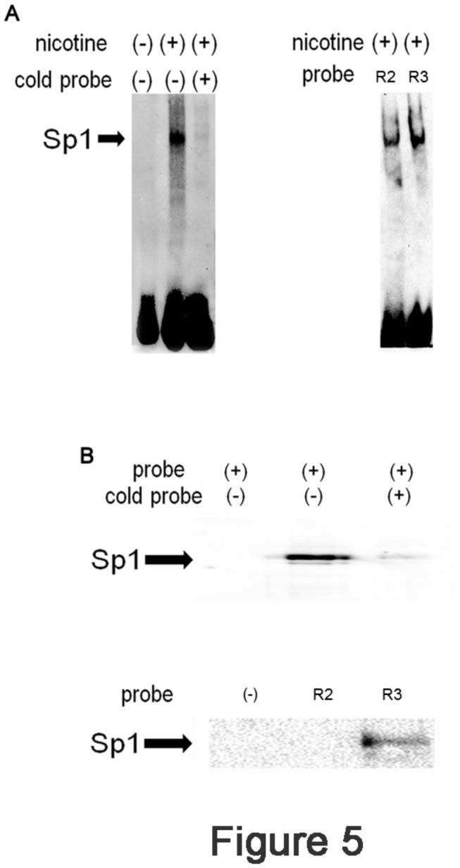
Figure 5. Nicotine stimulation leads to Sp1 binding to R3.

(A) Left panel: Ca9-22 cells were stimulated with (+) or without (-) nicotine for 3 h. After stimulation, the cells were harvested and the nuclear extracts were prepared. Twenty μg of nuclear extract was incubated with biotinylated R3 probe with (+) or without (-) cold R3 probe. The samples were separated by native PAGE and transferred to a nylon membrane. The retarded band was detected by incubating the membrane with streptavidine-HRP followed by ECL. The representative of 3 separate experiments was shown. Right panel: Nicotine-stimulated nuclear extract was incubated with or without R2 or R3 probe and the retarded bands were detected as described above. (B) Nicotine-stimulated nuclear extracts were prepared from Ca9-22 cells and subjected to Stre-Av precipitation assays. Upper panel: lane 1: Stre-Av precipitation assay performed without nuclear extract. Lane 2: Stre-Av precipitation assay with nuclear extract. Lane 3: Stre-Av precipitation assay with biotinylated and non-labeled R3 probes. Lower panel: lane 1: Stre-Av precipitation assay performed without probe. Lane 2: Stre-Av precipitation assay with R2 probe. Lane 3: Stre-Av precipitation assay with R3 probe.
