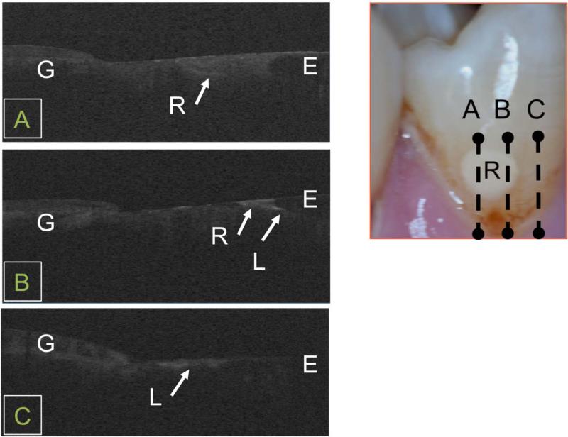
Fig. 6.
In vivo CP-OCT b-scans taken at three positions on a tooth (A, B, C ) with a restoration and an enamel lesion. The full extent of the restoration (R) can be seen including decay peripheral to the restoration (L) along the DEJ. An enamel lesion (L) can also be seen on the same tooth at position C.

