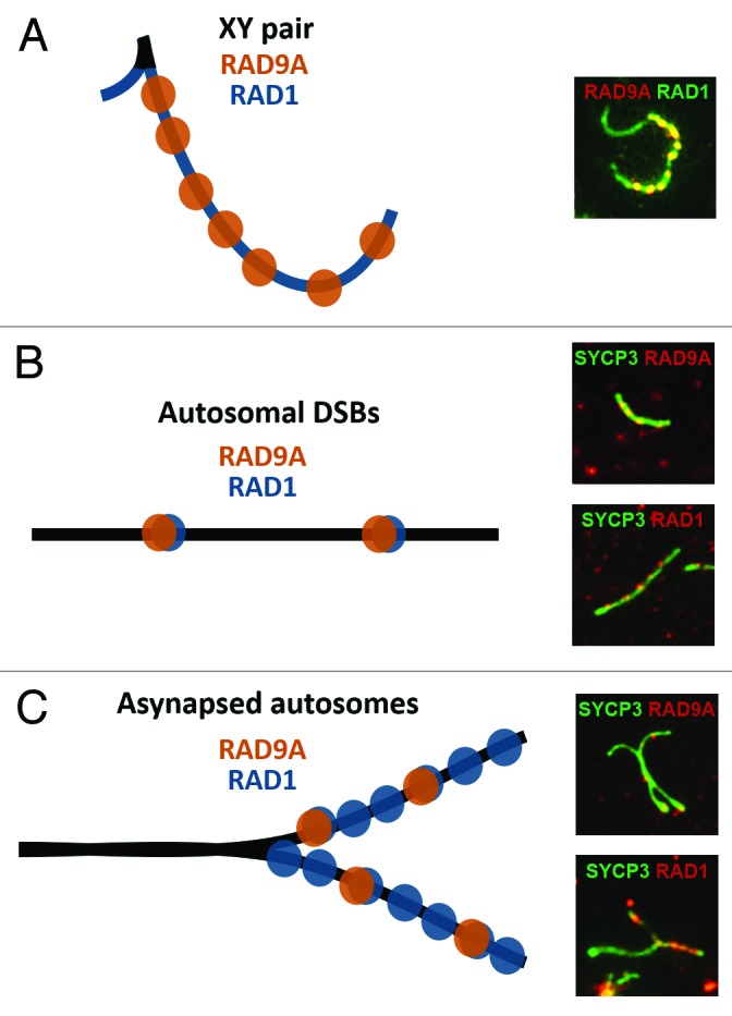
Figure 1. RAD9A and RAD1 proteins localize in distinct yet overlapping patterns on mammalian meiotic chromosomes. The localization patterns of RAD9A and RAD1 are shown in schematic form (left) and in representative immunofluorescence images (right).(A)During late pachynema, unsynapsed regions of the X and Y axial elements are continuously coated with RAD1, while the X chromosome axial element additionally harbors discrete RAD9A foci, presumably at sites of DSBs.(B)On early pachytene-stage autosomes, colocalization of RAD9A and RAD1 is observed in a focal pattern, likely marking DSB sites.(C)Asynapsed regions of autosomes show extensive RAD1 staining and discrete RAD9A foci. Similar to what occurs on the unsynapsed X and Y (A), RAD9A is present at sites that also contain RAD1, whereas RAD1 displays a broader staining pattern that includes regions without detectable RAD9A. See reference 15 for additional examples of these staining patterns.
