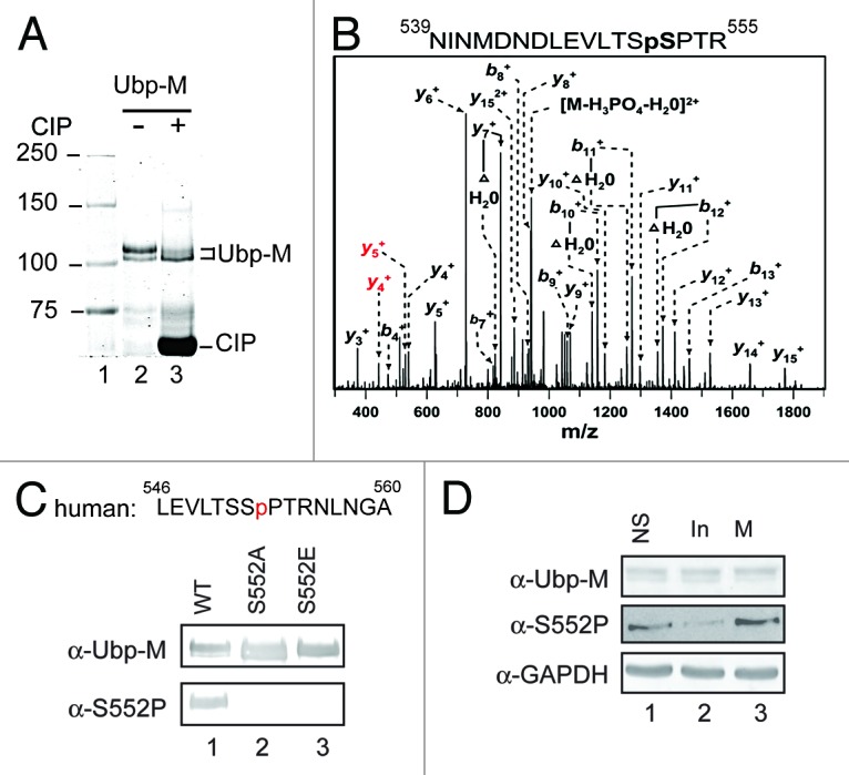
Figure 1.

Ubp-M is phosphorylated at the serine 552 residue. (A) Ubp-M is phosphorylated in vivo. Coomassie blue staining of affinity purified Ubp-M from sf9 cells incubated with (lane 3) or without (lane 2) Calf intestinal alkaline phosphatase (CIP). CIP treatment decreases the intensity of the top band and increases the intensity of the bottom band. (B) The serine 552 residue of Ubp-M is phosphorylated. Mass spectrometry analysis indicated that Ubp-M is phosphorylation at serine 552. (C) Generation of the Ubp-M phosphorylated serine 552 (S552P) antibody. Top: peptide sequence used to generate the Ubp-M S552P antibody. Bottom: characterization of the Ubp-M S552P antibody. Antibodies used are labeled on the left side of the panel. (D) Ubp-M S552P occurs in cell cycle M phase. Western blot analysis of extracts from unsynchronized, interphase, and M phase cells. Antibodies used are labeled on the left side of the panels.
