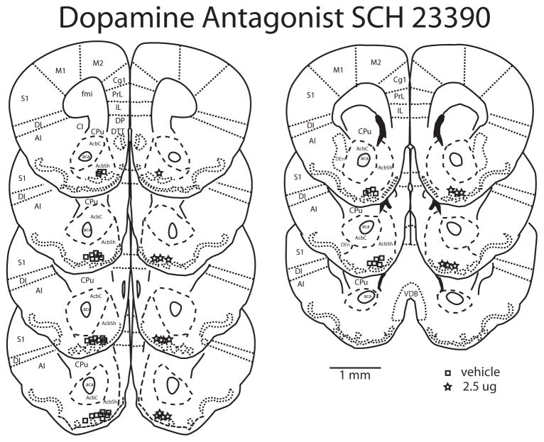
Figure 2.
Reconstructions of a coronally cut series of sections through the nucleus accumbens showing the histological verification of injection placement of D1 antagonist SCH 23390 into the nucleus accumbens shell. AcbC nucleus accumbens core; AcbSh nucleus accumbens shell; aca anterior commissure; AI agranular insula; DI dysgranular insula; S1 primary somatosensory cortex; Cgl cingulate cortex; PrL prelimbic cortex; M1 primary motor cortex; M2 secondary motor cortex; IL infralimbic cortex; DP dorsal peduncular cortex; DTT dorsal tenia tecta; fmi forceps minor or the corpus callosum; CPu caudate putamen; cl claustrum; DEn dorsal endopiriform nucleus; VDB vertical limb of diagonal band nucleus. All images are original work of the authors drawn from California mouse sections. Injection sites are represented by squares for vehicle and stars for 2.5μg SCH 23390. Scale bar is 1 mm.

