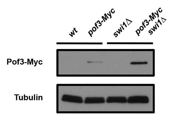
Figure 3. Pof3 is elevated in swi1∆ cells. Cells of the indicated genotypes were grown, and protein samples were prepared. Cellular amounts of Pof3-Myc were determined by using the anti-Myc (9E10) antibody. Tubulin levels were monitored as a loading control.
