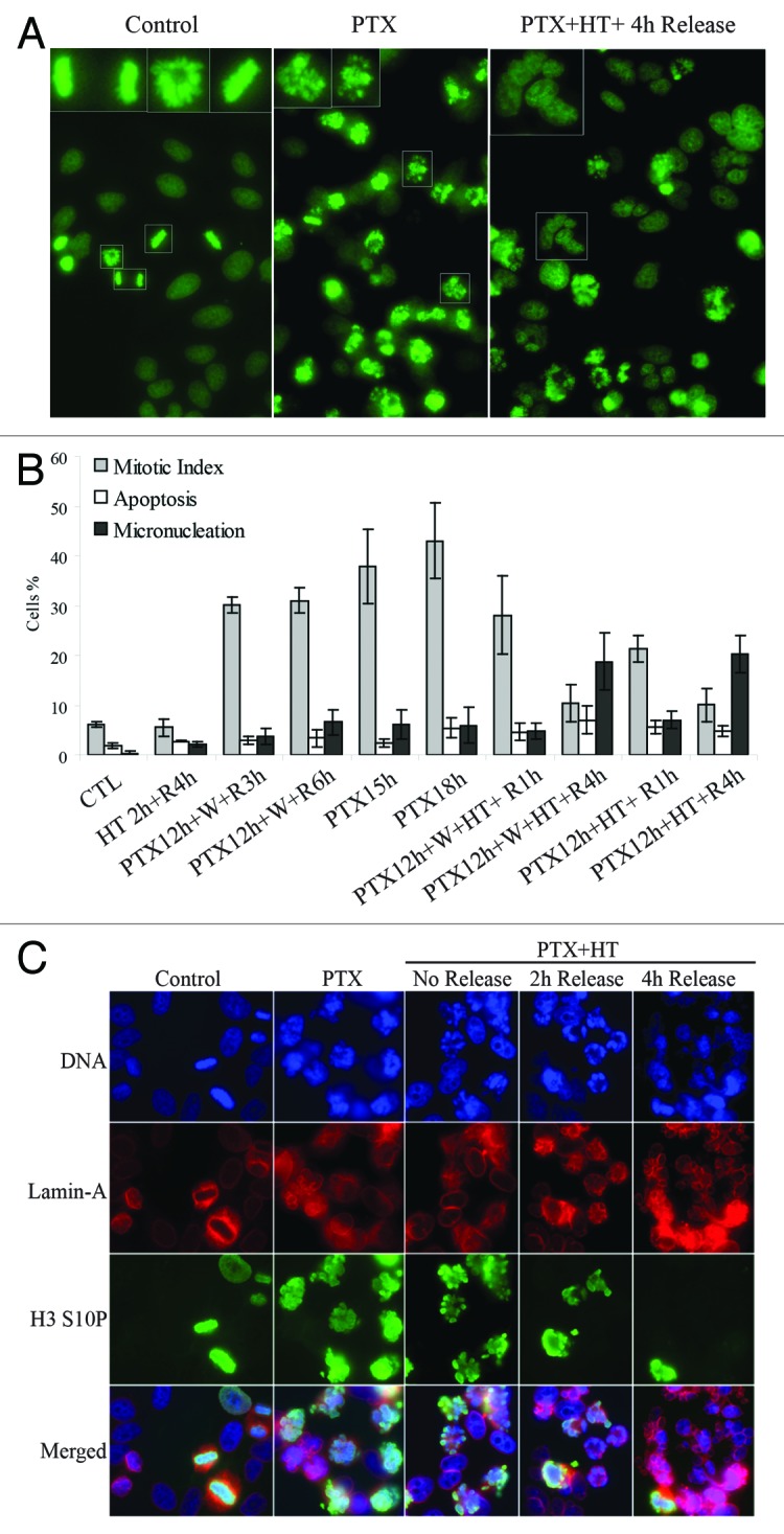
Figure 1. HT forces mitotic catastrophe in PTX-arrested cells. (A) DNA staining of HEp2 cells (control, left) treated with 10 nM PTX for 18 hours (h) at 37 °C (PTX) or treated with PTX at 37 °C for 12 h followed by a 42 °C HT for 2 h and returned at 37 °C for 4 h (PTX + HT + 4 h release). (B) Cellular morphology was analyzed to study the effects of PTX and HT. PTX treatment: 15 or 18 h, HT: 2 h, 42 °C; R, release at 37 °C for 1–6 h; W, PTX washout. At least 500 cells per sample were counted, (SD ± 3). HT forces mitotic exit and induces micronucleation in PTX-arrested cells. (C) Detailed analysis of HT-induced mitotic catastrophe. Immunoflourescence staining of HEp2 cells treated as in (A); Lamin A, marker of nuclear envelope; H3 S10P, mitotic marker H3 phospho-serine 10.
