
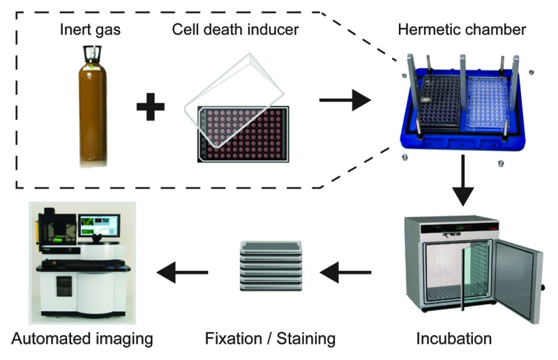
Figure 1. Experimental setup employed in this study. Human osteosarcoma U2OS cells stably expressing a histone 2B-red fluorescent protein (H2B-RFP) chimera were maintained in control conditions or exposed to a panel of cytotoxic compounds within gas tight chambers containing a standard atmosphere (20% O2, 5% CO2, and 75% N2) or a gas mixture in which N2 was specifically replaced with He, Ne, Ar, Kr, Xe, or N2O, while O2 and CO2 concentrations were kept unaltered. After 6 or 16 h, cells were fixed in the presence of Hoechst 33342, optionally stained for the detection of cytochome c release and caspase-3 activation, and subjected to robotized (immuno)fluorescence microscopy followed by automated image acquisition.