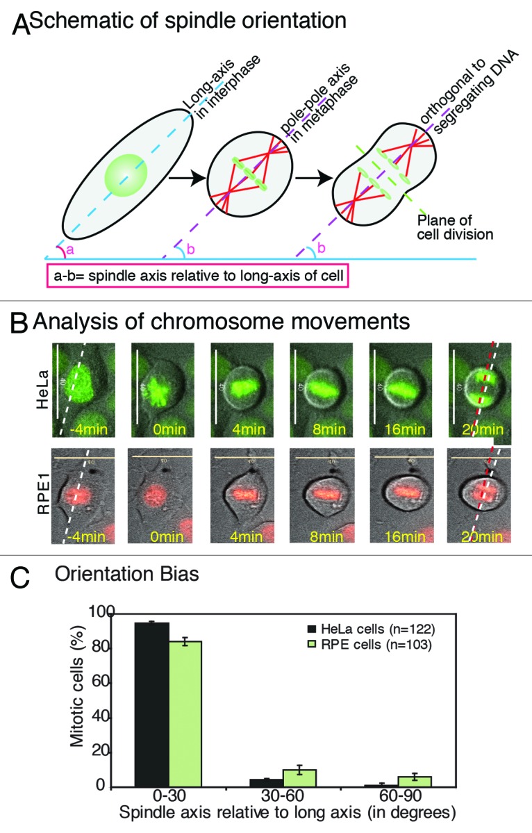
Figure 1. Epithelial cells cultured on glass orient their spindle along the interphase long-axis (A) Schematic describing cell shape-associated spindle orientation. The interphase long-axis (angle a; dashed blue line), metaphase spindle-axis (angle b; dashed purple line) and plane of cell division (dashed green line) orthogonal to spindle-axis are highlighted. (B and C) Spindle-axis at metaphase–anaphase transition shows biased alignment along the long-axis of the interphase cell. (B) Time-lapse analysis of chromosome movements to extract long-axis of the cell (dashed white line), spindle-axis (dashed red line) in HeLaHis2B-GFP and RPE1His2B-RFP cells. Representative frames of time-lapse movies are shown as a merge of DIC (cell shape) and either GFP/RFP (chromosome alignment) (C) Frequency distribution of final orientation angles (spindle-axis relative to long-axis) in HeLaHis2B-GFP and RPE1His2B-RFP. Error bars represent SEM from 3 independent experiments. Scale bar = 40 μm
