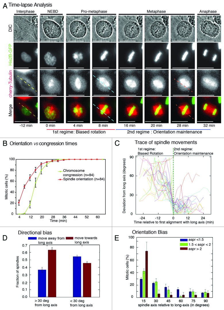
Figure 2. Two spatially distinct regimes of spindle movements facilitate the process of cell shape-dependent spindle orientation (A) Time-lapse images of a HeLaHis2B-GFP; mCherry-Tub cell showing the positions of spindle and chromosomes from nuclear envelope breakdown (NEBD) through anaphase. Dashed lines are drawn parallel to the interphase long-axis (yellow), first alignment along long-axis (blue), and final spindle orientation axis (pink). Scale bar = 15 μm. (B) Cumulative frequency distributions of the first time point when spindle-axis aligns with long-axis and the time point of completion of chromosome congression. Only cells that properly oriented along long-axis were considered for the analysis. (C) Trace of spindle-axis through time of a randomly chosen set of 20 cells. The vertical axis measures the angle between the spindle and the cell’s long-axis (0 corresponds to perfect alignment), each curve has been horizontally shifted, so that the time of first alignment (< 20 degrees) is at time 0. Dashed green vertical line indicates spindles’ first alignment with long-axis. (D) Bar graph showing direction bias for spindles rotating away from and toward the long-axis. Spindles were segregated into 2 sets according to their position (> or < 30 degrees from long-axis). (E) Frequency distribution of final orientation angles (spindle-axis relative to long-axis) in HeLaHis2B-GFP; mCherry-Tub cells that exhibited high (>2), medium (between 1.5 and 2) or low aspect ratio (<1.5) in interphase, 20 min prior to NEBD. Aspect ratio (aspr) is defined as major axis:minor axis of ellipse fitted to interphase cell shape, 20 min prior to NEBD. Error bars represent SEM from 3 independent experiments.

An official website of the United States government
Here's how you know
Official websites use .gov
A
.gov website belongs to an official
government organization in the United States.
Secure .gov websites use HTTPS
A lock (
) or https:// means you've safely
connected to the .gov website. Share sensitive
information only on official, secure websites.
