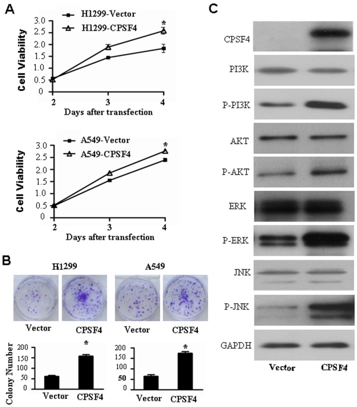
Figure 5. Overexpression of CPSF4 promotes cell growth and activates PI3K/AKT and MAPK signaling pathways in H1299 cells.
(A) H1299 cells were transfected with CPSF expressing vector, at 3 or 4 days cell viability was measured by MTT assay. The data are shown as the mean ± SD of three independent experiments (*, P<0.05). (B) H1299 cells were transfected with CPSF4 expressing vectors or control vector every 3 days and grown for 8 days, the colonies were stained with crystal violet and counted. The data are shown as the mean ± SD of three independent experiments (*, P<0.05). (C) H1299 cells were transfected with CPSF expressing vector, at 72 hours after transfection, the expression of CPSF4 as well as total and phosphorylated Akt, PI3K, ERK1/2 and JNK proteins was detected by Western blot. GAPDH was used as the loading control.

