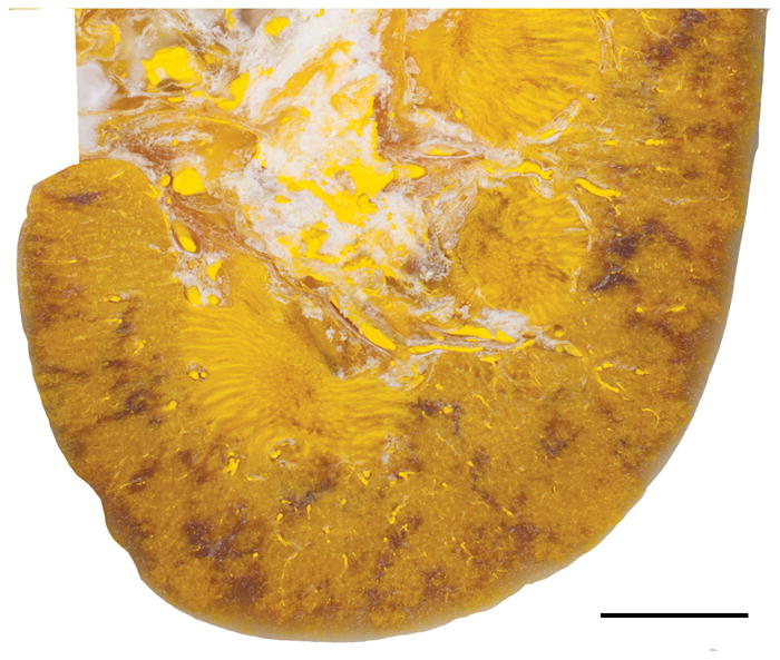
FIG. 1.

Light photograph of a cross-section of kidney from a pig treated with ultrasonic propulsion. Note the lack of hemorrhagic lesion after treatment to displace stone/beads using simulated clinical treatment parameters. The yellow color of the tissue is due to Microfil infusion of the vasculature used to mark the location of blood vessels to help distinguish vessels from tissue hemorrhage. The diffuse non-yellow regions of the cortex and medulla represent areas of imperfect Microfil infusion. Bar = 1 cm.
