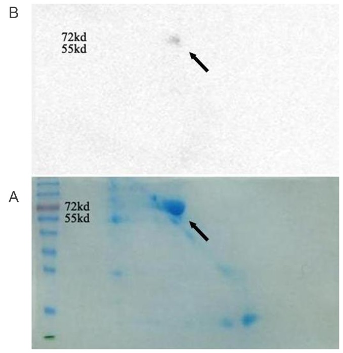
Figure 2. SDS-PAGE in two dimensions and Western blot analysis of seminal vesicle fluid shows a molecular weight 55-72KDa spot (arrowhead) binded to Eppin.
A. Seminal vesicle fluid was separated by 2-D electrophoresis\ and was stained overnight with 0.01% Bio-Rad R-250 Coomassie in 10% acetic acid.
B. Far Western blot: Incubate the membrane with 6-His-Eppin protein and probed with anti-His. We can see the positive spot molecular weight 55-72KDa on the blot (arrowhead).

