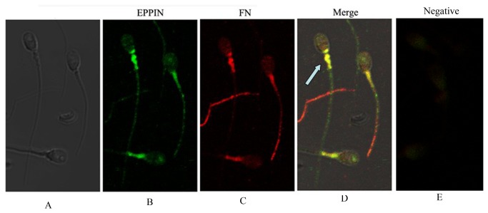
Figure 5. Immunolocalization of Fn and eppin in the sperm.
A Phase contrast image of spermatozoon.
B. Immunolocalization of Eppin on the head and tail of ejaculate human spermatozoa with anti-Eppin antibodies and detected with fluorescein isothiocyanate (FITC)-conjugated goat anti-rabbit antibodies (green).
C. Immunolocalization of Fn on the head and tail of ejaculate human spermatozoa with anti-Fn antibodies and detected with Cy3 (cyanine-3)-conjugated goat anti-mouse antibodies (red).
D. Dual-immunofluorescence staining of Fn (red) and Eppin (green) in human sperm showing that the strong colocalization (→) is seen in the postacrosomal and midpiece region of the head (yellow areas).
E. Control exposure with fluorescein isothiocyanate (FITC) conjugated goat anti-rabbit IgG and Cy3 (cyanine-3)-conjugated anti-mouse IgG, with isotype mouse antibody and rabbit antibody instead of primary antibody.

