
Figure 2.
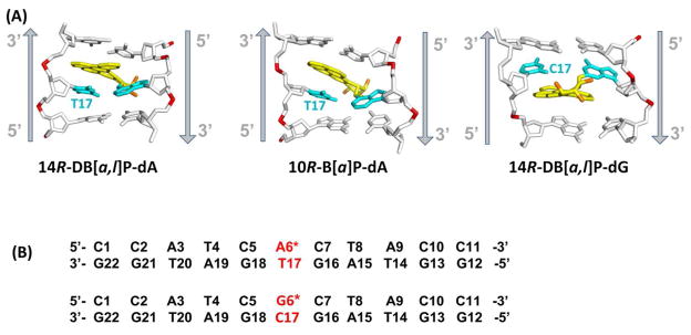
(A) Best representative structures49 from MD simulations for the 14R-DB[a,l]P-dA, and 10R-B[a]P-dA adducts. The NMR solution structure for the 14R-DB[a,l]P-dG adduct is also shown.42 Only the central 3-mers are presented. The view is looking into the minor groove. For the DB[a,l]P and B[a]P moieties, the carbon atoms are colored yellow and the oxygen atoms orange. The damaged bases are colored cyan and the DNA duplexes are colored white, except for the phosphorus atoms, which are red. Hydrogen atoms are not displayed for clarity. The arrows indicate 5′ to 3′ direction. Movies S1 – S3, showing these structures rotating are provided in Supporting Information. (B) The base sequence contexts for the 14R-DB[a,l]P-dA and the 14R-DB[a,l]P-dG 11-mer duplexes. A* and G* designate the damaged bases.