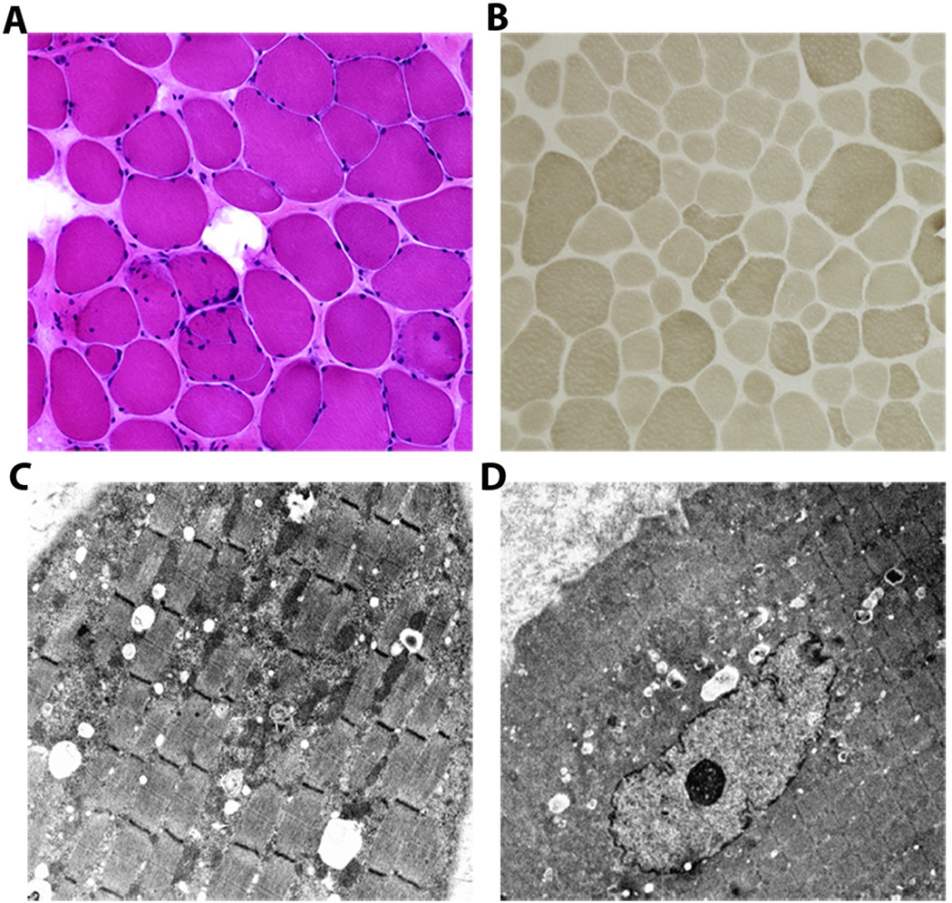
Figure 4. Morphological findings of quadriceps muscle biopsy sections from affected individual OH IV:1.
(A) Hematoxylin and eosin stain showing variability of fiber sizes with increased endomysial connective tissue and some internalized nuclei (20X). (B) Myosin adenosine triphosphatase (ATPase) 9.4 stain demonstrating fibers of variable sizes with small type I (light) and II (dark) fibers (10X). Electron micrographs show focal accumulation of mitochondria accompanied by glycogen and lipid droplets (C; 4000X); with some fibers showing an internalized nucleus (D; 2500X).

