
Figure 7. Expression of TIMPs.
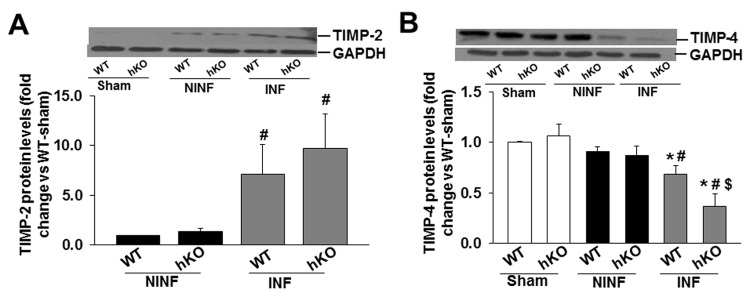
Total LV lysates (50 µg), prepared from sham and non-infarct (NINF) and infarct (INF) LV regions, were analyzed by western blot using anti-TIMP-2 (A) and anti-TIMP-4 (B) antibodies. The upper panels show autoradiograms indicating immunostaining for TIMP-2, TIMP-4, and GAPDH. The lower panels exhibit quantitative analyses of TIMP-2, TIMP-4 normalized to GAPDH. *p<0.05 vs sham; #p<0.05 vs NINF; $p<0.05 vs WT-INF; n=7.