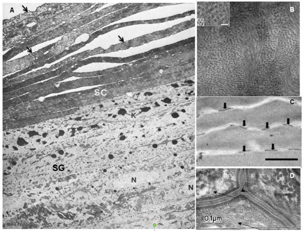
Figure 2. Transmission electron micrographs illustrating unique features of skin.
(A) The stratum corneum (SC) is comprised of multiple layers of enucleated and elongated corneocytes each defined by a dense cornified envelope indicated by the black arrows. The cells in the stratum granulosum (SG) are distinguished by the presence of a nucleus (N) and a high density of keratohyalin granules (K). Adapted with permission from Madison et al., 1998 [24] (B) Electron micrograph of a corneocyte cytosol illustrating the nanoscale organization of keratin intermediate filaments. The subfilamentous molecular architecture appears as groups of electron dense spots surrounding a central dense dot. The keratin filaments are ~7.8 nm wide with a center-to-center distance of ~16 nm. (Inset box scale bar is 10 nm. Adapted with permission from Norlen and Al-Amoundi, 2004 [25] (C) Corneocytes in the stratum corneum are bound by corneodesmosome tight junctions indicated by black arrows. Racial differences exist in the density of corneodesmosomes. Scale bar is 1 μm. Adapted with permission from Gunathilake et al., 2009 [27]. (D) Electron micrograph illustrating the lipid lamellar bilayers in the intercellular space between corneocytes. Adapted with permission from Warner et al., 1999 [30].

