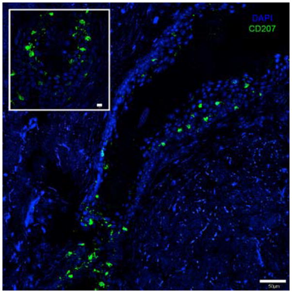
Figure 3. Langerhans Cell localization pattern around the hair follicle.
Immunofluorescent staining of human skin epidermis with anti-CD207-Alexa 488 (Langerin) specific for Langerhans cells showing their distribution around the hair follicle infundibulum, scale bar=50 μm. Inset shows the base of the hair follicle, scale bar=10 μm.

