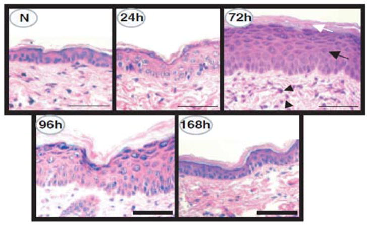
Figure 7. Characterization of UVR-induced epidermal injury in SKH-1 mice.
Hematoxylin and eosin stain photomicrographs of normal dorsal mouse skin (N) and of skin 24, 72, 96, or 168 h after UVR (180 mJ per cm2 UVB) irradiation. Note the epidermal hyperplasia (black arrow), hyperkeratosis (white arrow), and the perivascular inflammation (arrowheads) present 72 h after UVR irradiation. At 168 h post UVR irradiation, the epidermis has returned to near normal. Scale bar: 50 μm. Reprinted with permission from Tripp et. al., 2003 [80].

