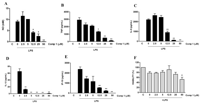
Figure 3. Compound 1 inhibits the production of pro-inflammatory mediators induced by LPS in macrophages.
Peritoneal macrophages were treated with the indicated concentrations of compound 1 (2.5, 5, 12.5, 25 or 50 μM). After 1 hour cells were stimulated with 1 μg/mL (A) or 10 ng/mL (B-F) of LPS. Supernatants were collected 24 h after the stimulus and NO (A), TNF-α (B), IL-6 (C), IL-1β (D) and IP-10 (E) concentrations were determined. (F) Cell viabilities were assessed using a MTT assay after supernatant collection. Results represent means ± S.E.M. from stimuli performed in duplicates and are representative of three different experiments. *, P ˂ 0.05; **, P ˂ 0.01, compared with LPS stimulus alone. (F) *, P<0.05 compared with the control without stimulus.

