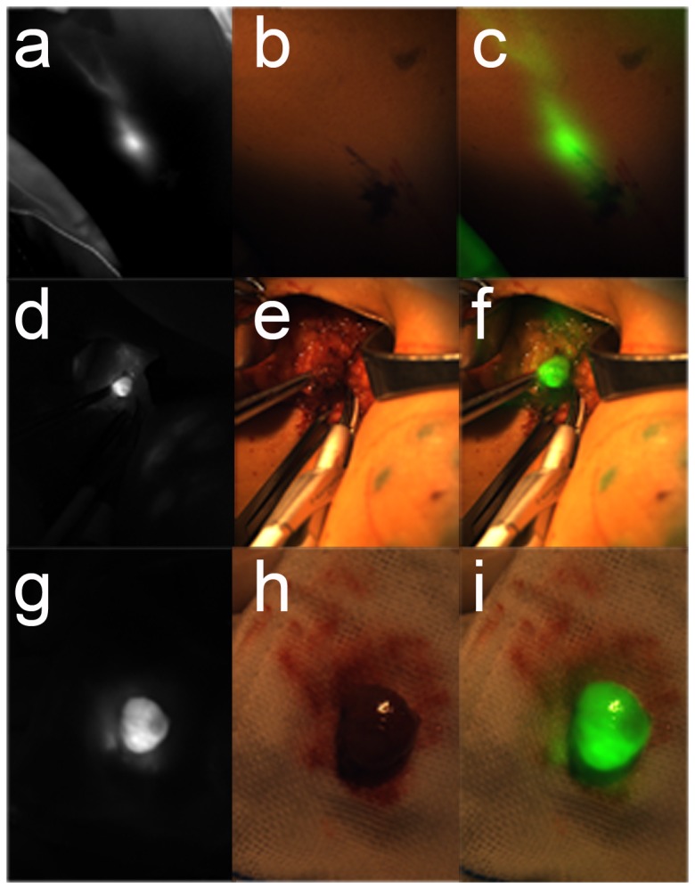
Figure 5. ICG-guided intraoperative detection and resection of the SLN in humans.

According to the preclinical trials, 22 cases of patients were taken from the SLNB surgery. In the beginning, the ICG solution was injected into the areolar region. About 3 minutes later, the lymphatic drainage and SLN would be clearly displayed on the monitor as shown in a. the fluorescent image in vivo. Because near-infrared light is not visible, there was no light information in the b. color image in vivo. Through the software of the surgical navigation system, the location of the SLN is shown in c. where the overlay image in vivo could be distinguished accurately. According to the guidelines of the fluorescent image, the surgery could quickly find the location of the SLN. d. The fluorescent image was captured before dissection. From the e. color image and the f. overlay image, SLN could be located with tweezers. The SLN was carefully removed and put on gauze. With the near-infrared light irradiation, the SLN was bright as shown in g. the fluorescent image during dissection. Such a visible image was displayed in h. the color image during dissection. Finally, the merged image of the pseudo-green fluorescence image and the color image is shown in i. the overlay image during dissection. All dissections were sent in for pathological examination. All of the tissue sections were judged to be the SLN.
