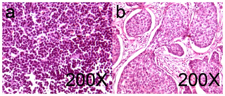
Figure 6. The normal and tumor-metastasis pathology slices of SLN dissected by ICG-guided surgery.

All of the dissected SLNs were sent in for pathological examination. After the conventional Hematoxylin-Eosin (HE) staining, the results proved that all of the dissected tissue specimens were lymph nodes. Figure a. shows normal sentinel lymph node cells with no cancer metastasis. Figure b. shows infiltrating ductal breast cancer.
