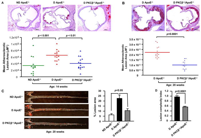
Figure 2. Impact of PKCβ deletion on diabetic atherosclerosis.
Shown are representative images of aortic root sections stained with Oil Red O at age 14 weeks (A) and 20 weeks (B) and Sudan IV stained aortic en face at age 20 weeks (C). Scale bar =200 μm. Mean atherosclerotic lesion areas (μm2) were determined in ND ApoE−/− (n=13), D ApoE−/− (n=14) and D PKCβ −/−/ApoE−/− male mice (n=15 and n= 13) at age 14 weeks (A) and 20 weeks (B). Lesion complexity index was calculated in D ApoE−/− and D PKCβ −/−/ApoE−/− male mice at age 20 weeks (n=10 and n=13) (D). D: diabetic; ND: nondiabetic.

