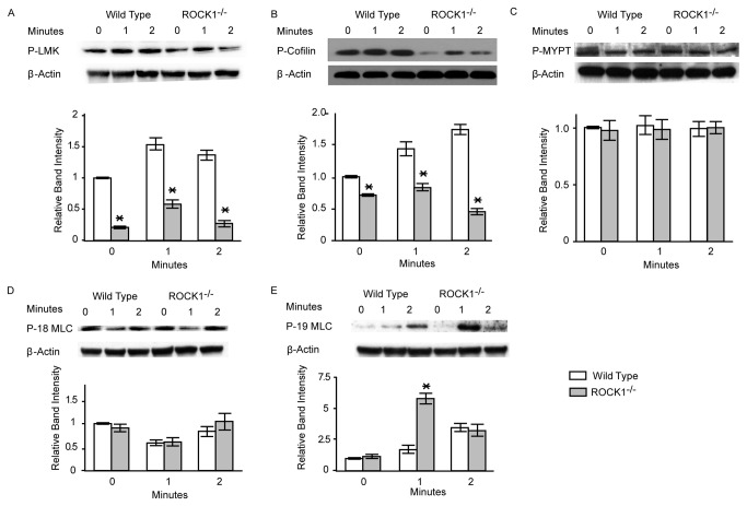
Figure 6. ROCK1-deficieny results in reduced phosphorylation levels.
Isolated platelets from ROCK1−/− and wild-type mice were stimulated with collagen (10 µg/ml). Platelets were solubilized at the indicated time intervals, subjected to SDS-PAGE and immunoblotted with antibodies against phospho-Lim Kinase-1 (LMK), phospho-cofilin-1, phospho-myosin phosphatase target subunit-1 (MYPT1); phospho-myosin light chain (threonine 18, P-18 MLC), and phospho-myosin light chain (serine 19; P-19 MLC). Immunoblot of β-actin was used as a loading control. One representative blot and group data (n= 3/group) are shown. * denotes a P value of less than 0.05 compared to wild-type.

