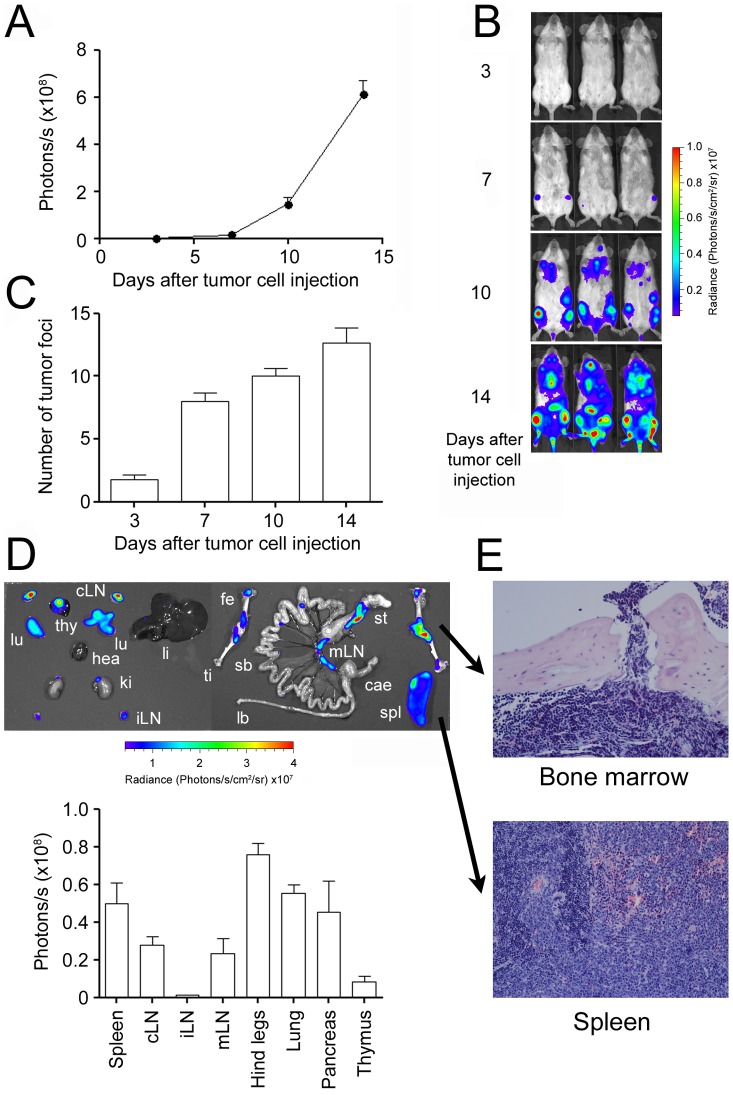
Figure 2. Non-invasive assessment of in vivo tumor growth and dissemination.
A: 105 luciferase-transgenic IM380 tumor cells were injected i.v. into the lateral tail vein into syngeneic BALB/c mice. Tumor growth was assessed by non-invasive in vivo BLI at the indicated time points. B: Representative BLI pictures of tumor-bearing mice. C: Tumor dissemination was determined by counting individual light-emitting tumor foci. D: Upper panel: Representative ex vivo BLI picture of a tumor bearing mouse (lu: lung, cLN: cervical lymph nodes, thy: thymus, hea: heart, ki: kidney, iLN: inguinal lymph nodes, li: liver, fe: femur, ti: tibia, sb: small bowel, lb: large bowel, mLN: mesenteric lymph nodes, st: stomach, cae: caecum, spl: spleen). Lower panel: Evaluation of tumor cell infiltration in individual organs. A–D: (Mean ± SEM; n = 5; shown is one representative experiment out of two). E: Representative eosin and hematoxylinstainings of organs from tumor bearing mice shown in 200× magnification.

