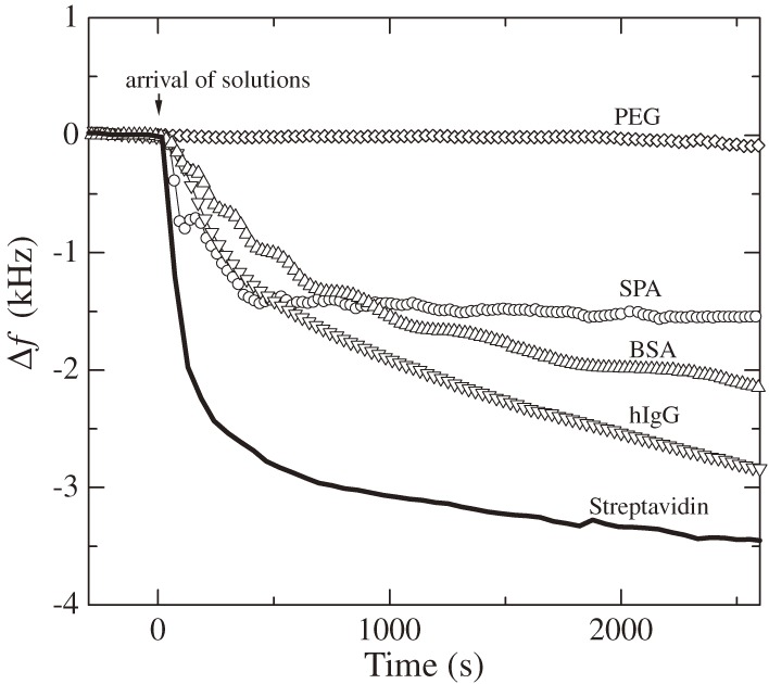
Figure 5.
Frequency changes observed for injections of 100 µg/mL concentration proteins in a phosphate buffered saline (PBS) solution (f1 = 55 MHz). PEG, SPA, BSA, and hIgG represent polyethylene glycol, staphylococcus protein A, bovine serum albumin, and human immunoglobulin G, respectively. This figure was reproduced with permission from Ogi et al.31)

