Figure 1.
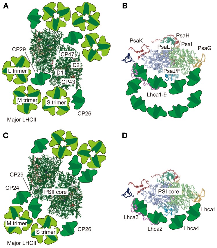
Supramolecular organization of PSII-LHCII and PSI-LHCI supercomplexes in green algae and vascular plants. Top views of the PSII-LHCII supercomplex (A) and the PSI-LHCI supercomplex (B) from C. reinhardtii based on single-particle image analysis by Tokutsu et al. (2012) and Drop et al. (2011), respectively. Top views of the PSII-LHCII supercomplex from spinach (C) and the PSI-LHCI supercomplex from pea (D) based on single-particle image analysis by Dekker and Boekema (2005) and crystallography of the PSI-LHCI supercomplex (Amunts et al., 2010), respectively. All top view images are from the lumenal side. The PSII and PSI core structures were taken from the coordinates determined by crystallography in 3ARC.pdb and 2WSC.pdb, respectively.

