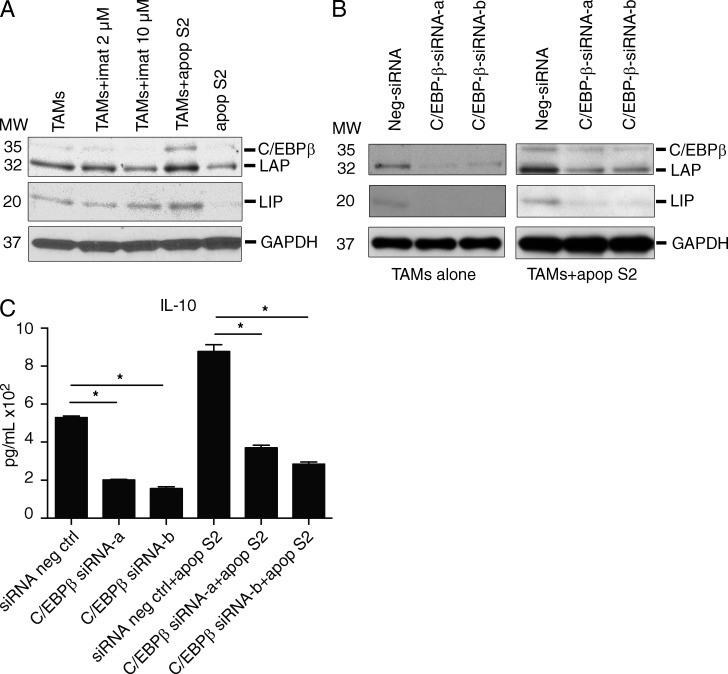
Figure 5.
Apoptotic tumor cells induce an M2-like shift via C/EBPβ. (A) S2 cells were rendered apoptotic by irradiation with 20 Gy and treatment with imatinib for 3 h and then washed to remove imatinib. 106 apoptotic S2 cells (apop S2) were then cultured with 2 × 10⁶ TAMs for 48 h in 6-well plates. Some wells were treated with 2 or 10 µM imatinib (imat). After 48 h, cell lysates were analyzed by Western blot. Shown is a representative of two experiments. MW, molecular weight. (B and C) 106 TAMs were transfected with either control (siRNA neg ctrl) or C/EBPβ-targeted siRNA (constructs a or b) for 24 h and then cultured with or without apoptotic S2 cells (apop S2). After 48 h, cell lysates were analyzed by Western blotting (B) and supernatant IL-10 was measured by cytometric bead array (C). Shown is a representative of two experiments. Bar graphs show mean ± SEM. *, P < 0.05.

