Figure 3.
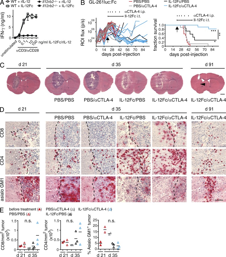
Local administration of IL-12Fc in established GB in combination with systemic CTLA-4 blockade induces tumor clearance. (A) IFN-γ production of WT or Il12rb2−/− splenocytes stimulated with anti-CD3 and anti-CD28 mAbs and increasing amounts of heterodimeric IL-12 (rIL-12) or IL-12Fc; one out of three experiments shown. (B) WT mice were injected i.c. with GL-261luc:Fc cells. On day 21 after injection, osmotic minipumps delivering IL-12Fc or PBS intratumorally (i.t.) were implanted as indicated by a horizontal line above graphs. In addition, animals received i.p. injections with αCTLA-4 mAbs or PBS, indicated by vertical tick marks; n = 7–12/group; three independent experiments pooled. Log-rank test; *, P < 0.05; **, P < 0.01; ***, P < 0.001; p(IL-12Fc/PBS vs. IL-12Fc/αCTLA-4) = 0.0061, p(PBS/PBS vs. IL-12Fc/PBS) = 0.0160, p(PBS/PBS vs. IL-12Fc/αCTLA-4), and p(PBS/αCTLA-4 vs. IL-12Fc/αCTLA-4) < 0.0001. (C–E) Histological analysis of GL-261luc:Fc tumors at day 21, 2 wk after initiation of treatment (day 35) and at the end of the experiment (day 91). (C) Overviews, arrowhead indicates former tumor center. Bar, 2 mm. (D) Higher magnification, sections show CD4, CD8, and asialo GM1 immunoreactivity (red); tumor margin is indicated (dotted line. Bar, 200 µm. (E) Quantification of TILs/tumor area on histological sections, each dot represents mean of a single animal; one-way ANOVA with Bonferroni post-test; one of two independent experiments shown; *, P < 0.05; **, P < 0.01; ***, P < 0.001.

