Figure 2.
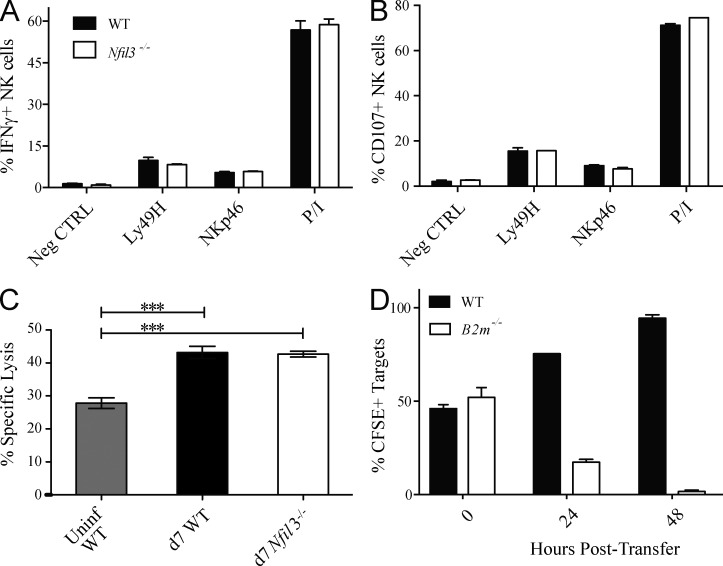
Comparable function of WT and Nfil3−/− NK cells after MCMV infection. Splenic WT and Nfil3−/− NK cells were stimulated for 5 h with anti-Ly49H, anti-NKp46, or PMA + Ionomycin (P/I) and evaluated for IFN-γ production (A) and degranulation (B; as determined by CD107a expression) by flow cytometry. (C) 51Cr-labeled YAC-I target cells were incubated with splenic WT or Nfil3−/− NK cells isolated at day 7 PI. Specific lysis was determined 4 h later. (D) CFSE-labeled WT and B2m−/− targets were injected (i.v.) into MCMV-infected Nfil3−/− mice at day 7 PI, and specific killing (percentage of CFSE+ cells remaining) was evaluated at the indicated time points after transfer. Error bars for all graphs show SEM and data are representative of three independent experiments with n = 3 mice per group.

