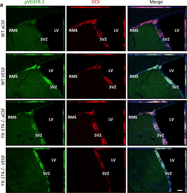
Figure 7.
Phosphorylation of VEGFR-2 is increased in NPCs of the SVZ of Flt-1TK−/− mice and mice that intracerebrally received VEGF-A. Sagittal sections of the brain showing VEGFR-2 phosphorylation (green) in DCX+ cells (red) counterstained with DAPI (blue) in the aSVZ of the LV. VEGFR-2 is phosphorylated particularly in DCX+ cells of aSVZ and the RMS. The intensity of phospho-VEGFR-2 immunoreactivity is much greater in the aSVZ and RMS of Flt-1TK−/− mice and in mice that received VEGF-A compared with WT mice. Scale bar, 200 μm.

