
Figure 3.
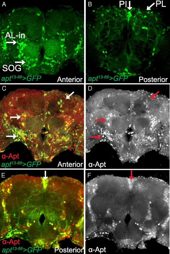
Apt expression in the adult CNS. A, B, Whole-mount brain from an adult male fly apt13–66-GAL4/UAS-GFP (nls);UAS-mCD8GFP/+ stained with anti-GFP. Confocal projections are from the anterior (A) and posterior (B) regions of the brain. A, In the anterior brain, apt13-66-GAL4 expression (green) is found in AL-in and neurons in the SOG. B, In the posterior brain, apt13–66-GAL4 expression is observed in neurons of the PL and PI. C–F, Whole-mount brain from an adult male fly apt13–66-GAL4/UAS-GFP (nls);UAS-mCD8GFP/+ stained with anti-GFP (green) and anti-Apt (red). Projections are from the anterior (C, D) and posterior (E, F) regions of the brain. C, E, There is colocalization of apt13–66-GAL4 (green) and Apt immunoreactivity (red) in the AL-in, SOG, PL, and PI (yellow; white arrows). D, F, Anterior and posterior projections showing the expression pattern of Apt in the Al-in, SOG, PL, and PI (red arrows).