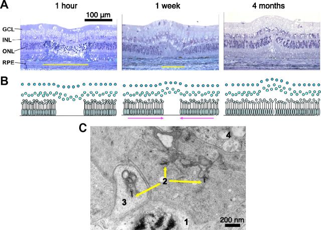
Figure 1.
Restoration of retinal anatomy after photocoagulation. A, Toluidine-blue-stained retinal sections at various time points after the “barely visible” grade photocoagulation. Yellow bars show the lateral extent of the photoreceptor layer damage. B, Diagrammatic representation of the restoration of the photoreceptor layer continuity. C, TEM photograph of a central area of a healed 2-month-old lesion showing: (1) the photoreceptor nucleus, (2) photoreceptor ribbon synapses, (3) synaptic vesicles, and (4) bipolar cell processes.

