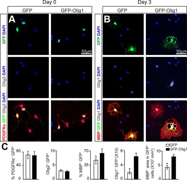
Figure 7.
Overexpression of Olig1 in Olig2-deleted OPCs in vitro rescues differentiation arrest. A, B, OPCs from Olig2cnp-cko mice were purified and transfected with Olig1-GFP or GFP constructs and immunostained the next day after plating (0 d) for Olig1 (white), PDFGRα (red) (A) or cultured in OPC differentiation media for 3 d and stained for Olig1 (white) and MBP (red) (B). C, Quantification of the immunostaining demonstrates that OPCs transfected with the Olig1-GFP plasmid promote the expansion of MBP+ membrane area compared with OPCs transfected with GFP alone without changing the number of PDGFRα+ cells. Scale bar: A, B, 50 μm. Data are mean ± SEM, and all experiments were performed in triplicate. *p < 0.05.

