Figure 2.
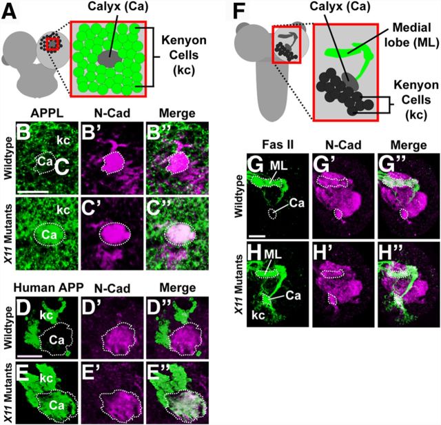
X11 is required in the MB for polarized localization of human APP, APPL, and fasciclin II. B–E”, G–H”, Staining of brains against Drosophila N-cadherin (N-Cad, magenta) strongly labels the calyx of the MB, which is outlined by a dotted line, allowing for its identification. A, An illustration depicts polarized localization of endogenous APPL (green) in Kenyon cells (kc) and the calyx (Ca) of a wild-type MB, viewed posteriorly. B–B”, Wild-type first instar brains stained with anti-APPL antibody indicate APPL (green) is excluded from the MB's calyx. C–C”, In contrast, brains of X11 mutants stained against APPL show mislocalization of APPL to the calyx and increased levels in Kenyon cells. Scale bar: (in B) B–C”, 10 μm. D–D”, Similarly, third instar brains exogenously expressing myc-tagged human APP695 by MB-specific driver OK107-GAL4 and stained with anti-Myc antibody indicate ectopic human APP (green) is mostly somatoaxonally localized, with very low levels detectable in the calyx. E–E”, However, expression of RNAi transgenes against X11 causes human APP to be dramatically increased in Kenyon cells and the calyx. Scale bar: (in D) D–E”, 50 μm. B–E”, All brains stained against human APP or APPL were imaged from the posterior side. F, Illustration depicts polarized localization of endogenous fasciclin II (green) in a wild-type MB, viewed dorsally. G–G”, Wild-type first instar brains stained with anti-fasciclin II (Fas II) antibody indicate Fas II (green) is excluded from the calyx and Kenyon cells of the MB. H–H”, Brains of X11 mutants stained against Fas II show mislocalization of Fas II to the calyx and Kenyon cells, indicating loss of polarized localization. G–H”, Brains stained against Fas II were imaged from the dorsal side. Mislocalization of Fas II is also visible in brains of X11 mutants imaged from the posterior side. Scale bar, 10 μm.

