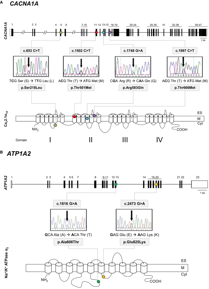
Figure 2.

Gene structure of CACNA1A and ATP1A2 and detection of mutations. (A) 1: CACNA1A gene structure, with black boxes indicating exons. The identified mutations causing HM are indicated by colored dots: p.Ser218Leu (light green), p.Thr501Met (red), p.Arg583Gln (blue), and p.Thr666Met (purple). 2: Protein structure and location of the identified mutations. (B) 1: ATP1A2 gene structure, with black boxes indicating exons. The newly identified mutation p.Glu825Lys causing HM is indicated by a yellow dot and p.Ala606Thr with a green dot. 2: Protein structure and location of the identified mutations. Detection of the mutations by direct sequencing of PCR products: electropherograms. Cyt, cytoplasm; M, cytoplasmic membrane; ES, extracellular space.
