
Fig. 4.
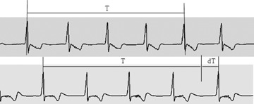
Effects of gastric distention on gastric slow wave frequencies in a dog. Top: 80-sec. slow wave recordings from a pair of serosal electrodes at the baseline. Bottom: 80-sec. slow wave recordings from the same pair of serosal electrodes when the stomach was distended by an intragastric balloon with a pressure of 40 mmHg. An obvious decrease in slow wave frequency can be appreciated by comparing the time intervals covering four cycles of slow waves at the baseline (T) and during distention (T + dT): the longer time period for the four slow waves during distention is indicative of a decrease in the slow wave frequency.