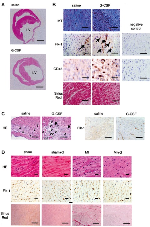
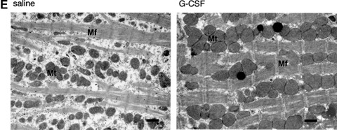
Fig. 1.


Effect of G-CSF on cardiac morphology 4 weeks after MI. (A) Transverse sections of infarcted hearts. Masson's trichrome staining. Bars, 1 mm. (B) Histological (Masson's trichrome, Sirius red) and immunohisto-chemical (Flk-1, CD45) staining of the infarcted area of a saline-treated and G-CSF-treated heart. The most right panels show negative control sections for immunostains. Arrows indicate immunopositive cells. Bars, 50 μm in CD45 staining; 100 μm in the others. (C) Haematoxylin and eosin and Flk-1 immunohistochemical staining of the border zones between infarcted and non-infarcted area of a saline-treated and G-CSF-treated heart. Arrows, arterioles; arrowheads, venules. Bars, 100 μm. (D) Histological staining (haematoxylin and eosin and Sirius red) of the non-infarcted area of hearts. Bars, 100 μm. (E) Ultrastructure of salvaged cardiomyocytes. Mf, myofibrils; Mt, mitochondria. Bars, 1 μm.
