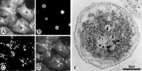
Fig. 3.

Fluorescence and electron microscopy of MPIO-labelled primary human hepatocytes. Cells were stained against cytokeratine 18 (A) and nuclei (B). Dragon Green labelled MPIOs (C) were distributed throughout the cytoplasm. In the overlay (D), cytokeratine 18 is illustrated as red, nuclei as blue and MPIOs as green. Electron microscopy of single hepatocytes proved the intracellular localization of the particles, both as single particles and as clusters (E).
