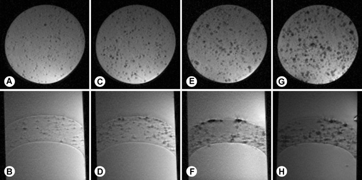
Fig. 4.

Agarose samples of hepatocytes with increasing particle load were investigated using T2*-weighted, two-dimensional gradient echo pulse sequence by 3.0 Tesla in sagittal and axial slices. Cells containing 10 ± 2 particles/cell (A, B) or 16 ± 1 particles/cell (C, D) showed areas of slight hypo-density. Preparations of cells containing 18 ± 1 particles (E, F) or 25 ± 2 particles (G, H) displayed punctuate signal extinctions that clearly contrast against the high-signal image background caused by the aqueous agarose medium. Imaging parameters were as follows: repetition time/echo time/flip angle = 200 msec/25 msec./20°. Spatial resolution was 78 μm × 78 μm × 800 μm requiring scan times of 5.10 min.
