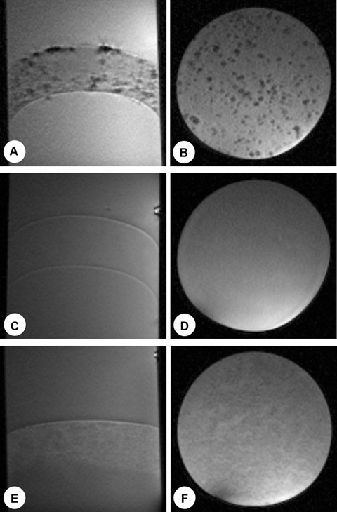
Fig. 5.

Cells labelled with 18 ± 1 MPIOs/cell were clearly detectable both in sagittal and axial slices (A, B) at a concentration of 1000 cells/250 μl. The same number of non-labelled cells (C, D) or a correlating number of MPIOs suspended in agarose (E, F) showed no detectable distinct signal changes. Imaging parameters were the same as in Fig. 4.
