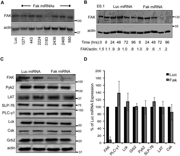
Figure 1.
MicroRNAs repress FAK expression in human T cells. A, Jurkat cells were transfected with either a control (Luc) miRNA or various miRNAs specific for FAK. Whole cell lysates were taken 72 h later and the expression of FAK and actin was analyzed by immunoblotting. B, Jurkat T cells were transfected with Luciferase- or FAK-specific miRNAs. Whole cell lysates were prepared at the indicated times and FAK and actin expression were analyzed by immunoblotting. The densitometric ratio of FAK expression compared to actin expression is shown below the graph. C. Whole cell lysates from Jurkat E6.1 T cells transfected with microRNAs against Luciferase or FAK were analyzed by immunoblot for FAK, Pyk2, LAT, SLP-76, PLC-γ1, Lck, Csk, and Grb2 and actin expression 72 h after transfection. (D) The normalized expression of FAK, Pyk2, LAT, SLP-76, PLC PLC-γ1, Lck, Csk, or Grb2 expression is shown as the mean of two to three experiments ± s.d.

