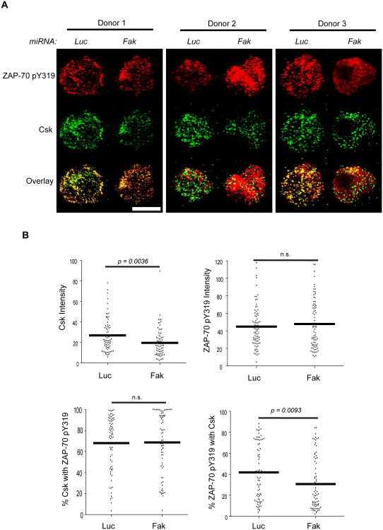
Figure 6.
Csk membrane and TCR localization is altered in FAK-deficient CD4 hAPBTs. A, CD4 hAPBTs were transfected with the Luc or FAK-specific microRNAs and incubated for 72 h. The cells were stimulated for 7.5 min with anti-TCR coated onto glass chamber slides. These cells were then stained with anti-phospho-ZAP70 Y319 and anti-Csk followed by the appropriate secondary antibodies. Representative cells from three separate human donors are shown. The white scale bar is equal to 5 μm. B, The intensity of Csk and ZAP-70 pY319 staining and co-localization between these proteins was analyzed as in Figure 9. The values from 75 cells taken from three independent experiments are shown and the mean values are represented by the horizontal lines. The p-values reflect the statistical differences between the control and FAK-deficient cells at the indicated times. n.s. means p > 0.05.

