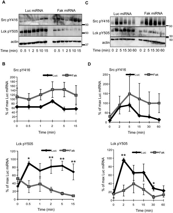
Figure 8.
The phosphorylation of Lck is altered in FAK-deficient human T cells. A, Luciferase (Luc) or Fak miRNA-containing Jurkat T cells were stimulated as in Figure 2. The site-specific phosphorylation of Src Y416 (equivalent to Lck Y394) and Lck Y505 was then analyzed by immunoblotting. B, Quantification of three to four experiments examining the phosphorylation of Lck in control and FAK-deficient Jurkat cells. Data is shown as mean ± s.e.m. C, Control or Fak-deficient primary human CD4 T cells were stimulated as in Figure 3. D, Percent phosphorylation of Src Y416 and Lck Y505 in stimulated control or FAK-depleted primary human CD4 T cells was determined. Data is depicted as mean of three experiments ± s.e.m. * p ≤ 0.05; ** p ≤ 0.01.

