
Fig. 3.
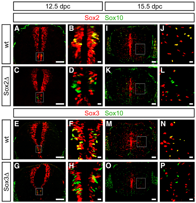
Efficiency of Sox2 and Sox3 deletion in Sox2ΔSox10 and Sox3ΔSox10 oligodendroglia. Co-immunohistochemistry on transverse sections of wild-type (wt) (A,B,E,F,I,J,M,N), Sox2ΔSox10 (Sox2Δ) (C,D,K,L) and Sox3ΔSox10 (Sox3Δ) (G,H,O,P) SC at 12.5 dpc (A-H) and 15.5 dpc (I-P) with antibodies against Sox2 (A-D,I-L) or Sox3 (E-H,M-P) (red) and Sox10 (A-P) (green). Higher magnifications of framed areas from A,C,E,G,I,K,M,O (scale bars: 100 μm) are shown in B,D,F,H,J,L,N,P (scale bars: 10 μm).