Fig. 3.

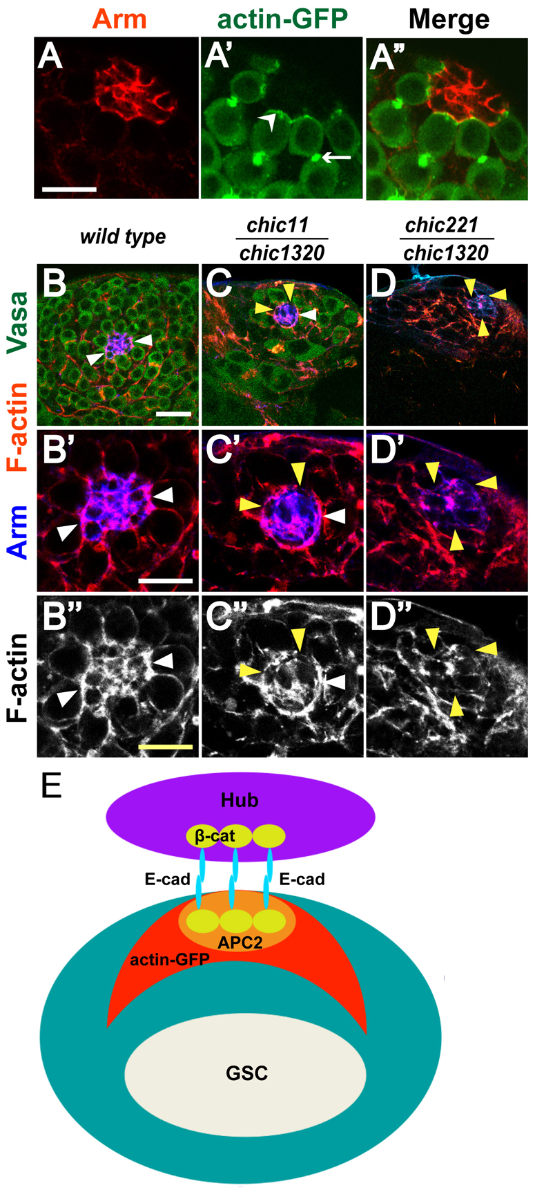
F-actin is enriched at the hub-GSC interface. (A-A′) Localization of actin-GFP in GSCs. Adult testis tip from a nanos-GAL4; UAS-actin-GFP fly stained with (A) anti-Arm (red), (A′) anti-GFP to reveal actin-GFP expression in germ cells. (A′) Merge. Arrowhead indicates GSC-hub interface. Arrow indicates fusome. Scale bar: 10 μm. (B-D′) Larval testis tips from (B-B′) wild type, (C-C′) chic11/chic1320 hypomorph and (D-D′) chic221/chic1320 strong loss-of-function animals with anti-Vasa (green) to mark germ cells, phalloidin to highlight F-actin (red) and anti-Arm (blue) to mark hub cells. White arrowheads indicate F-actin localized to hub-GSC interface. Yellow arrowheads indicate loss of F-actin at the hub perimeter. Scale bars: 20 μm in B-D; 10 μm in B′-D′. (E) Localization patterns of adherens junction components: E-cadherin and β-catenin, APC2 and actin-GFP.
