
Fig. 1.
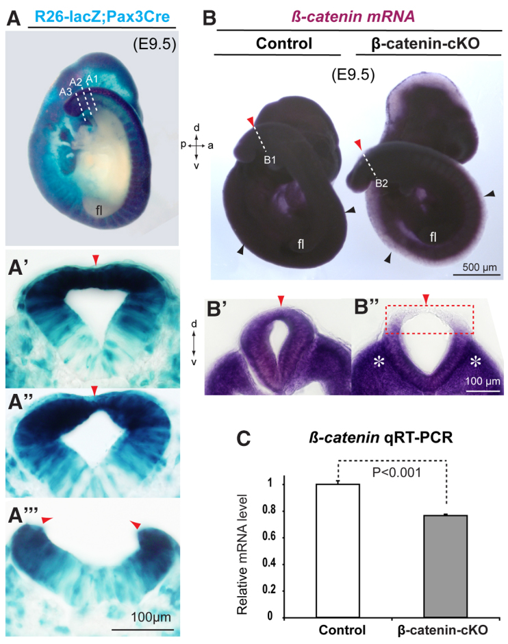
Effective ablation of β-catenin in the mouse dorsal neural tube with Pax3-Cre. (A-A′′) X-gal staining (blue) for genetic fate mapping of Rosa26-lacZ;Pax3Cre/+ demonstrates the Cre recombination pattern in the dorsal neural tube, including closed (section A1 in A′), closing (section A2 in A′) and pending closure (section A3 in A′′) posterior neuropore (PNP). (B-C) In situ hybridization and real-time PCR results for conditional inactivation of β-catenin mRNA in the dorsal neural tube of the β-catenin cKO [the abbreviation for β-catenin(ex2-6)flox/flox;Pax3Cre/+] at E9.5. Sections B1 and B2 are shown in B′ and B′, respectively. Note that β-catenin mRNAs were ablated in the dorsal PNP (boxed region in B′), but were still expressed in the majority of the cKO tissues including the ventral neural tube and the paraxial mesoderm (asterisks in B′) around the PNP closure site. Error bars indicate s.e.m. (n=3). Black arrowheads, dorsal neural tube midlines; red arrowheads, the dorsal midline around the PNP closure regions; dashed lines, planes of transverse PNP sections. a, anterior; d, dorsal; fl, forelimb bud; p, posterior; v, ventral.