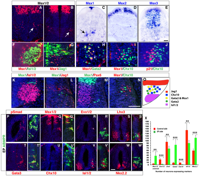
Fig. 5.
BMP/TGFβ signaling promotes V2b cell fates at the expense of V2a interneurons in the ventral spinal cord. (A-N) Msx expression analysis in the ventral spinal cord. Cells immunoreactive for Msx1/2 were observed in the ventral spinal cord of E10.5 mouse embryos (arrow in A) and stage 18 chick embryos (arrow in B). In situ hybridization on E11.5 mouse spinal cords revealed Msx1 expression in dorsal as well as ventral domains (C, arrow points to ventral Msx1 expression), Msx2 expression in a dorsal region (D), and Msx3 expression in a broad dorsal domain (E). In E10.5 mouse (F-J) and stage 20 chick (K-N) ventral spinal cords, no Msx1 expression was observed in ventrally located motoneurons that express Isl1/2 (F,K), dorsal cells that express Jagged1 (G,L), or in Pax6-positive progenitors (M). Msx1 overlaps with Gata2 in proximal Gata2-expressing cells, but is shut off in distal Gata2-expressing cells (H). It does not colocalize with Chx10 in V2a neurons (I,N). p21 expression, like Msx1, also precedes Chx10 expression in this domain (J). Transverse sections through the spinal cord are shown in A-N. (O) Schematic of expression patterns of Msx1 and other markers in the ventral spinal cord. (P-W′) Overexpression of ABMPR increased the number of pSmad-, Msx1/2-, Gata3- and Nkx2.2-immunoreactive cells on the electroporated side (P-Q′,T,T′,W,W′), but inhibited the number of Evx1/2-, Lhx3-, Chx10- and Isl1/2-immunoreactive cells (R-S′,U-V′). The asterisks in (R-S′,V,V′) indicate the exclusion of Evx1/2-, Lhx3- and Isl1/2-positive cells from the regions with overexpressed ABMPR (green). (X) Quantification of the effect of ABMPR overexpression on ventral cell fates. Each histogram represents the mean±s.d. for three embryos. Statistical significance was determined by Student’s t-test: **P<0.005, ***P<0.0001. Scale bars: in E, 10 μm for A,B; in E, 15 μm for C-E; in N, 37 μm for F,G; in N, 18.5 μm for H-J; in N, 20 μm for K-N; in W, 13 μm for P-W′.

