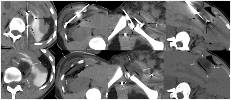
Figure 2.
Nonenhanced axial CT images matching the locations noted in Figure 1, showing cryoprobe in place (top row) and then removed (bottom row) to better define low-density ice (arrows) encompassing the prior tumor regions. All procedures were performed on an outpatient basis, with the patient discharged approximately 4 hours later.

